Research

Research

ASL MRI & CBF Mapping


Arterial spin labeling (ASL) is a non-invasive MRI technique that uses magnetically labeled blood water as an endogenous tracer to measure cerebral blood flow (CBF) and arterial transit time (ATT). The IRIS Lab has been a leader in advancing ASL acquisition and analysis, developing deep-learning methods — including 3D convolutional neural networks — to generate high-fidelity parametric perfusion maps from noisy multi-delay ASL data. The lab has also contributed to large-scale clinical trials such as the U.S.

AD Risk Neuroimaging


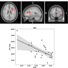
A central mission of the IRIS Lab is to understand how cerebrovascular and brain structural changes relate to cognitive decline and Alzheimer's disease (AD) risk. The lab's work — conducted in collaboration with Wake Forest School of Medicine, UC Davis, and partners in the U.S. POINTER lifestyle intervention trial — examines how metrics such as cerebrovascular reactivity (CVR), cerebral blood flow, white matter integrity, and arterial stiffness differ between individuals with normal cognition and those with mild cognitive impairment (MCI).

Deep Learning for Neuroimaging Analysis


The IRIS Lab actively develops and applies deep learning methods to solve challenging problems in neuroimaging, from image reconstruction to anatomical segmentation and parametric mapping. Notable contributions include a 3D convolutional neural network (CNN) that simultaneously estimates CBF and ATT from multi-delay ASL data, bypassing traditional fitting approaches that are sensitive to noise. 

WM & Vascular Health

White matter — the brain's network of myelinated axonal pathways — is highly sensitive to vascular insults, making it a critical imaging target for studying aging and disease. The IRIS Lab investigates the interplay between white matter microstructural integrity (measured via diffusion MRI), white matter perfusion (measured via ASL), and white matter hyperintensities (WMH), which are markers of small vessel disease visible on conventional MRI.

Sustainable MRI

Medical imaging systems — particularly MRI, CT, and PET/CT — are among the most energy-intensive pieces of equipment in a hospital, yet their consumption patterns are poorly understood at the suite level. The IRIS Lab investigates how to characterize, quantify, and ultimately reduce the energy footprint of these modalities. Using novel analytical frameworks such as load-duration curves, the lab maps moment-to-moment power draw across full imaging sessions and identifies opportunities for efficiency gains without compromising diagnostic quality.